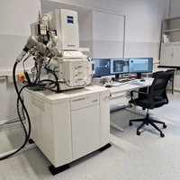
FIB-SEM, skaningowy mikroskop elektronowy SEM z działem jonowym FIB
FIB-SEM Crossbeam 350 z kolumną GEMINI i źródłem elektronów FEG oferuje obrazowanie w wysokiej rozdzielczości przy użyciu zaawansowanych trybów detekcji, w tym InLens (SE), InLens (EsB), Angle Selective Back-Scattered Detektor (AsB). Ustawienia obrazowania optyki Gemini , takie jak napięcie przyspieszające lub prąd wiązki, można płynnie regulować (z wybranym określonym skokiem). Równoległa detekcja elektronów wtórnych (SE) w osi soczewki obiektywowej oraz selektywne energetycznie rozproszenie wsteczne (EsB) pozwala z łatwością zidentyfikować najmniejsze różnice w składzie chemicznym materiałów. Wyposażony w zaawansowany system zogniskowanej wiązki jonów FIB, który umożliwia szybkie i precyzyjne usuwanie warstwy materiału oraz wycinanie próbek (lameli) dla FIB-SEM Crossbeam 350 z kolumną GEMINI i źródłem elektronów FEG oferuje obrazowanie w wysokiej rozdzielczości przy użyciu zaawansowanych trybów detekcji, w tym InLens (SE), InLens (EsB), Angle Selective Back-Scattered Detektor (AsB). Ustawienia obrazowania optyki Gemini II, takie jak napięcie przyspieszające lub prąd wiązki, można płynnie regulować. Równoległa detekcja elektronów wtórnych (SE) w osi soczewki obiektywowej oraz selektywne energetycznie rozproszenie wsteczne (EsB) pozwala z łatwością zidentyfikować najmniejsze różnice w składzie chemicznym materiałów. Wyposażony w zaawansowany system zogniskowanej wiązki jonów FIB, który umożliwia szybkie i precyzyjne usuwanie warstwy materiału oraz wycinanie próbek (lameli) dla TEM. Możliwości Crossbeam można rozszerzyć za pomocą oprogramowania Atlas 5 (ZEISS), wiodącego na rynku pakietu do szybkiej, precyzyjnej tomografii. Crossbeam 350 (ZEISS) ma optykę elektronową Gemini i optykę jonów FIB dostosowaną do szybkości i precyzji.
Na warunkach uzgodnionych z Kierownikiem laboratorium - dr hab. inż. Adam Kruk, prof. AGH
InLens (SE), InLens (EsB), Angle Selective back-scattered detector (AsB), Tomografia FIB-SEM
Obrazowanie mikrostruktury w elektronach wtówrnych SE i wstecznie rozproszonych BSE. Tomografia FIB-SEM. Wykonanie lameli do badań TEM.
Jednostka odpowiedzialna