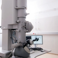
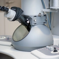
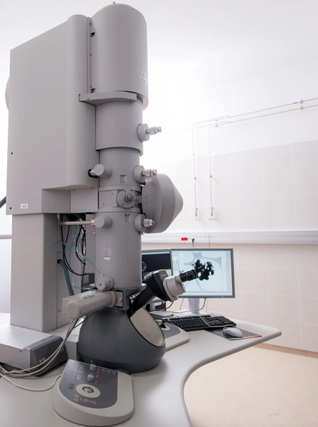
Transmisyjny mikroskop elektronowy (TEM) Tecnai TF 20 X-TWIN (FEI)
Tecnai TF 20 X-TWIN (200 kV) jest wysokorozdzielczym transmisyjnym mikroskopem elektronowym wyposażonym w działo elektronowe z emisją polową (Field Emission Gun - FEG). Mikroskop umożliwia obrazowanie mikrostruktury szerokiej gamy materiałów (takich jak: metale i ich stopy, ceramika, półprzewodniki, polimery, kompozyty) w jasnym i ciemnym polu (ang. bright field/dark field imaging). Obserwacje tego typu pozwalają na analizę m.in. wielkości ziaren/krystalitów w materiale, zdefektowania materiału, rozmieszczenia i rozkładu wielkości wydzieleń, grubości powłok, itp.. Wysoka zdolność rozdzielcza mikroskopu (punktowa zdolność rozdzielcza ≤ 0,22 nm, limit informacyjny ≤ 0,14 nm) umożliwia szczegółowe obrazowanie nanostruktur (nanocząstki, nanodruty, nanowarstwy, kropki kwantowe), jak również bezpośrednie obrazowanie struktury atomowej materiałów. Ponadto, za pomocą dyfrakcji elektronowej (dyfrakcja w wiązce równoległej, Selected Area Diffraction) istnieje możliwość: identyfikacji faz w konkretnych mikroobszarach w materiale, analizy lokalnej orientacji mikroobszarów, czy też określenia zależności krystalograficznych występujących między poszczególnymi fazami w materiale. Mikroskop posiada też zintegrowany spektrometr promieniowania rentgenowskiego (EDAX) służący do analizy składu chemicznego preparatów w mikro-/nanoobszarach. Mikroskop ma też możliwość pracy w trybie skaningowo-transmisyjnym (STEM) z detektorem obrazowym HAADF.
Mikroskop charakteryzują następujące parametry pracy:
- źródło elektronowe z emisją polową - FEG
- napięcie przyspieszające - 200 kV
- zakres powiększeń - 1025 – 900 k
- kamera CCD 2k (Eagle 2k HR)
- detektory: EDAX RTEM 0.3 sr, HAADF
Aparatura udostępniania na zasadach wynikających z Regulaminu Korzystania z Infrastruktury Badawczej ACMiN. (https://acmin.agh.edu.pl/home/acmin/5_Wspolpraca/Aparatura/Zasady_i_koszty_korzystania_z_infrastruktury_badawczej_ACMiN.pdf)
- Analiza morfologii i wielkości ziaren/krystalitów/cząstek, zdefektowania materiału, rozmieszczenia i rozkładu wielkości wydzieleń, grubości powłok, itp.
- Analiza struktury atomowej materiału (odległości międzypłaszczyznowe, identyfikacja defektów strukturalnych)
- Identyfikacja faz wchodzących w skład materiału, analiza lokalnej orientacji krystalograficznej mikroobszarów
- Analiza składu chemicznego w mikro-/nano- obszarach
- obserwacje w jasnym polu (BF), ciemnym polu (DF) oraz wysokorozdzielcze (HR)
- obserwacje w trybie skaningowo-transmisyjnym (STEM) z użyciem pierścieniowego detektora ciemnego pola (HAADF)
- dyfrakcja elektronowa z wybranych obszarów (SAED)
- analiza EDS (punktowa, liniowa oraz tzw. mapping).
Holdery: Double Tilt Low Background, Single Tilt
Oprogramowanie: TEM Imaging & Analysis, Low Dose, K-space control, Smart tilt


Jednostka odpowiedzialna
Grupa / laboratorium / zespół
Zakład Inżynierii Materiałowej