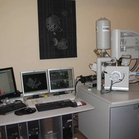
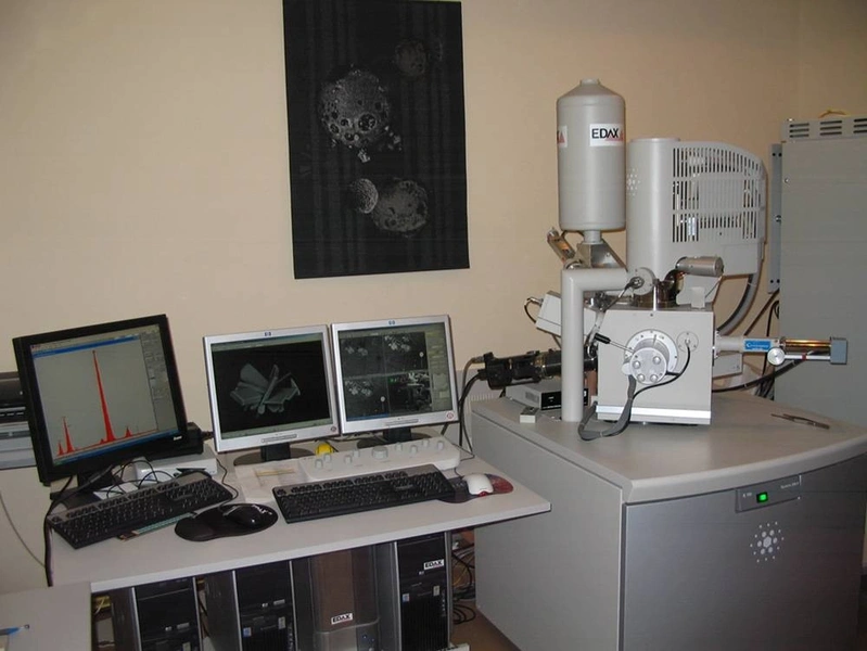
Mikroskop elektronowy skaningowy FEI Quanta 200 FEG
Wysokorozdzielczy skaningowy mikroskop elektronowy z gorącą emisją polową (FEG - wysokostabilny emiter elektronów Schottky'ego), zapewniający: możliwość obserwacji w zmiennej próżni, powiększenia 70 ÷ 300 000x, zdolność rozdzielczą ok. 2 nm.
Parametry techniczne:
- Napięcie przyspieszające 5 – 30 kV
- System zmiennej próżni:
- wysoka próżnia (HV) ok. 10-4 Pa - preparaty przewodzące
- niska próżnia (LV) 50÷200 Pa - preparaty przewodzące i nie przewodzące
- tryb ESEM 200÷2000 Pa - preparaty przewodzące i nie przewodzące
- Rozdzielczość:
< 2,0nm przy 30kV (HV)
< 3,5nm przy 30kV (LV)
< 2,0nm przy 30kV (ESEM)
Detektory:
- SED Everharta-Thornleya
- SED (do LV)
- SED (do ESEM)
- BSED (Si solid-state)
- BSED (do ESEM)
- Centaurus – fotopowielacz z końcówkami do detekcji BSE i CL
- EBSD (do rejestracji dyfrakcji elektronów wstecznie rozproszonych)
- Krzemowy, chłodzony ciekłym azotem, detektor promieniowania rentgenowskiego ze spektrometrem EDS (EDAX)
- zgodnie z regulaminem zamieszczonym na stronie laboratorium: http://wydzlab.agh.edu.pl/wp-content/uploads/2020/11/Regulamin-WLBFSTiG.pdf
- współpraca naukowo-badawcza z jednostkami AGH i innymi krajowymi i zagranicznymi jednostkami naukowymi
- współpraca w ramach projektów NCN, NCBiR i w ramach umów z przemysłem
- obserwacja powierzchni substancji mineralnych, syntetycznych i biologicznych
- analiza jakościowa i ilościowa pierwiastków w próbce (EDXS)
- analiza dystrybucji pierwiastków w mikroobszarze (EDXS)
- analiza obrazów w katodoluminiscencji CL - w trybie stopni szarości
- analiza dyfrakcji Kikuchiego (EBSD) - identyfikacja fazowa i mapy orientacji krystalitów OIM
Rejestracja, w zmiennej próżni, obrazów generowanych przez:
- elektrony wtórne SD – kontrast topograficzny
- elektrony wstecznie rozproszone BSE – kontrast różnicy Z i orientacyjny
- charakterystyczne promieniowanie rentgenowskie pierwiastków - mapowanie
- promieniowanie w zakresie widzialnym – katodoluminecencja CL
- EBSD - dyfrakcja Kikuchiego
Jednostka odpowiedzialna
Grupa / laboratorium / zespół
Wydziałowe Laboratorium Badań Fazowych, Strukturalnych, Teksturalnych i Geochemicznych